Month: August 2021

Liver diseases in Pregnancy

Liver diseases in pregnancy

These are the main type of liver diseases found during pregnancy-

AFLP – Acute fatty liver of pregnancy – characterized by maternal liver dysfunction usually in the third trimester, which can result in maternal or fetal complications, in some cases even fatal. It occurs due to a defect in the mitochondrial fatty acid beta-oxidation. Symptoms include nausea, vomiting, discomfort in the abdominal area and jaundice.

Preeclamptic liver dysfunction or preeclampsia induced liver disorder –This disorder is unique to pregnancy and occurs in the third trimesters. Preeclampsia can affect the liver and result in necrosis, hemorrhage that can cause nausea, vomiting, mild jaundice and abdominal pain. Severe form of preeclampsia can result in HELLP Syndrome – Haemolysis, Elevated Liver Enzymes and Low Platelet. It may also result in maternal or fetal death

ICHP Cholestasis: It is a liver problem where the flow of bile from the gall bladder decreases or stops. It may start in early pregnancy but is more common in the second and third trimester.  It can cause itching and the skin turn yellow. The pregnancy hormones may decrease the flow of bile. The bile made by the liver is stored in the gallbladder but if its levels keep increasing the bile will overflow in the liver and flow into the blood stream. High levels of bile can affect the fetus. This issue goes away soon after the delivery of the baby

Hyperemesis gravidarium: Liver disease occurs in around 50% of women diagnosed with HG. The severity of nausea and vomiting in HG is at times related to elevation of the liver (mild LFT – liver function test).


Symptoms of liver disease in pregnancy

  • Having Nausea or vomiting in the last semester can be a sign of Acute fatty liver or HELLP.
  • Yellow colouring of skin.
  • Puritis or Itching can be a symptom of ICH
  • Pain in the abdomen for some.
  • Abnormal liver tests
  • Gastrointestinal bleeding
  • Ascites – where fluid collects in the abdomen which may result in an infection

Complications of liver diseases

  • Jaundice
  • Disturbed sleep
  • Fetal distress
  • Preterm birth
  • Breathing problems
  • In severe cases maternal or fetal death.

Diagnosis for liver disease

The tests may vary depending on the disease the doctor suspects that the patient may be having. If the symptoms are in the

First trimester – Hyperemesis gravidarium

Second Trimester – ICH –usually 25 weeks or later

Third trimester – AFL in pregnancy and HELLP syndrome

Different tests include –

  • Liver function tests
  • Liver biopsy
  • Prothrombin time – time blood takes to clot
  • CBC and platelets
  • AST – aspartate aminotransferase
  • ALT – alanine aminotransfer
  • Bilirubin
  • Alkaline phosphatase
  • Albumin
  • Total protein
  • Basic panel
  • INR – International normalized ratio

Treatment for liver diseases during pregnancy

It is based on the type of liver disease identified, the stage of pregnancy and your health.

  • If you have been diagnosed with ALF they will suggest immediate delivery of baby else termination if it is in the early stage of pregnancy.
  • If diagnosed with ICH, treatment will be focused on reducing the itching and any related complications.
  • Steroid treatment for fetal lung development.
  • Early delivery – if you are at 37 or 38 weeks they may try to deliver the baby to avoid any further complications.
  • Continuous monitoring of the fetal growth.

For more details, kindly contact us.


Vaginal Infections/Vaginitis

vaginal infections

What are the methods of diagnosing vaginitis or vaginal infections in women?

Diagnosis of Vaginitis

Vaginitis includes a spectrum of conditions causing burning, itching, odour and vaginal discharge. Vulvo vaginitis is one of the commonest gynaecological conditions encountered in out patient clinics in UAE. Vaginitis can be caused by infections, allergy, hormone (Oestrogen) deficiency. The most common infectious causes of vaginitis are bacterial vaginosis, vulvo- vaginal candidiasis and trichomoniasis. Health care providers traditionally diagnose vaginitis using a combination of clinical symptoms, examination findings, pH of vaginal discharge, microscopy and special tests like ‘Whiff’ or ‘sniff’ test.

Diagnosis starts from the review of the medical history of vaginal or sexually transmitted diseases, next is to perform a pelvic examination and using a speculum to look into the vagina and collect a sample for lab testing and for wet mount and pH testing. An elevated pH is an indication of BV or TV. But pH alone is not reliable.

Bacterial vaginosis (BV) is caused by gardnerella vaginalis is the most prevalent cause of vaginal discharge. Bacterial vaginosis (BV) presents with malodorous, clear white or greyish discharge with a ‘fishy’ odour. Even when asymptomatic BV infection is having associated with pelvic inflammatory disease.

In our clinical practice, BV is diagnosed by the presence of 3 out of 4 Amsel criteria.

  • Thin homogenous vaginal discharge.
  • Vaginal pH greater than 4.5.
  • Positive whiff test (fishy odour or adding 10% potassium hydroxide solution to the vaginal discharge)
  • Atleast 20% of ‘clue cells’ (vaginal epithelial cells with borders observed by bacteria as wet mount preparation on a special stain named Gram’s stain’


In Trichomonias which is caused by protozoan presents with greenish yellow frothy discharge.

In vulvo-vaginal candidiasis caused by an yeast infection, presents with curdy white or cottage cheese like discharge and it can present with itchy, excoriations and swelling of the genital area.

Trichomoniasis – is diagnosed by microscope on wet mount preparation.

Candidiasis too is diagnosed by microscopic examination and one can visualize budding yeast in the wet smear of vaginal discharge.

What are the factors that increase the incidents of vaginitis in women?

  1. Sexual activity
  2. Hormonal changes like taking birth control pills, menopause and pregnancy
  3. Taking medication like antibiotics and steroids
  4. Uncontrolled diabetes
  5. Vaginal douching
  6. Wearing damp or tight under clothings
  7. Using intrauterine contraceptive device for birth control


What are the complications of vaginitis in women?

Women with TV or BV are at a greater risk of acquiring sexually transmitted infections. In pregnant woman, symptomatic BV and TV have been associated with premature birth and low birth weight babies even though there are conflicting studies about the same.

Bacterial vaginosis in pregnant women can predispose them to a slightly increased risk of preterm birth.

What are the ways to treat vaginitis?

Prevention and treatment:

Good personal hygiene is the cornerstone for the prevention of vaginitis.

Avoiding hot tubs and whirlpool spas etc. can help in preventing recurrent vaginitis especially in those who are prone for such infections.

Avoiding irritants like scented tampons, pads, douches and scented soaps and deodorants, etc. can be beneficial in prevention of these infections.

The basic practice of wiping from front to back after using toilet, avoid spreading of fecal bacteria to your vagina.

Other useful tips are to use cotton under wears.

BV is treated by anti bacterial medication which can be taken by mouth or vaginal gel. Yeast infections can be treated by anti-fungal creams and pessary and oral anti-fungal medications.

Trichomoniasis too can be treated by anti bacterial medication. Vaginal atrophy can be seen in menopause can be treated by oestrogen cream.


For more details kindly contact us.

Intrauterine growth restriction/Babies small for dates

IUGR

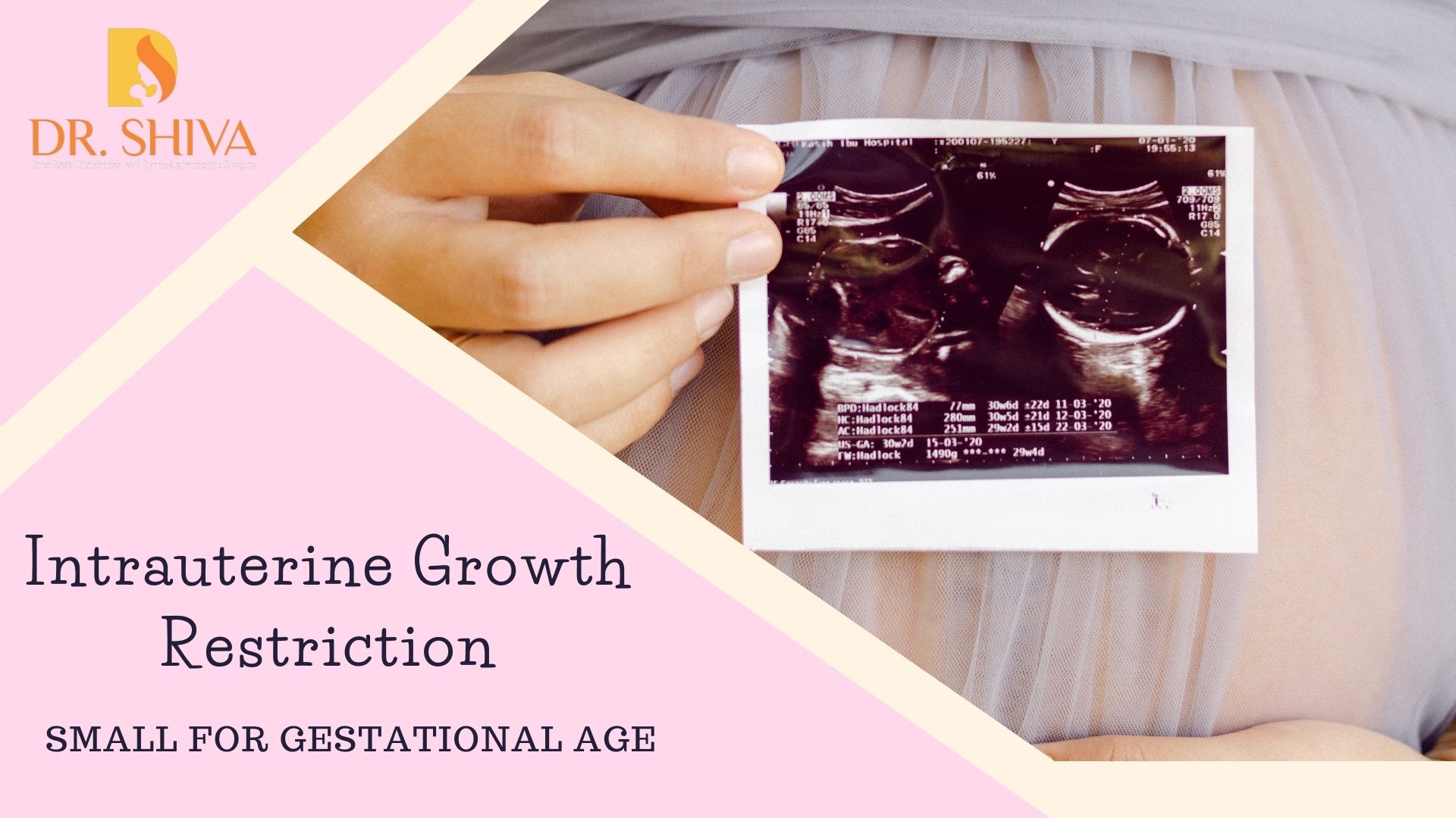
IUGR is when the fetus does not grow as expected or is smaller than it should be for that stage of the pregnancy. Mild IUGR is fine as the baby will grow to it’s appropriate size by 2 years but if it is severe it may cause serious health problems to the baby. The extent depends on the cause for it and at what point of pregnancy this has started.

They are of two types-

  • Symmetrical IUGR, when all parts of the body are of the same size but small.
  • Asymmetrical IUGR, when the head and brain are of expected size based on the month of pregnancy but the rest of the body is small.

Causes for IUGR

IUGR happens when the fetus is not getting the required amount of nutrients or oxygen. This can be due to,

  • an issue with the placenta which provides the nutrients to the baby.
  • a problem with the blood flow in the umbilical cord which connects the placenta to the fetus.

Other reasons include –

  • Usage of alcohol or drugs
  • Diseases like syphilis
  • High blood pressure
  • Kidney or lung disease
  • Autoimmune disease
  • Sickle-cell Anemia
  • Multiple babies – may result in an uneven share of blood and nutrients between the babies
  • Carrying a baby with birth or genetic defect

What are the risks associated with IUGR?

  • Babies with IUGR may have to stay at the hospital for a longer duration or at NICU if they were born early.
  • They may have an issue with breathing and feeding
  • low blood sugar levels
  • neurological problems
  • high count of red blood cells
  • low immunity
  • low oxygen levels
  • may also result in stillbirth

IUGR cannot be avoided but a healthy lifestyle can help reduce the risks associated with it.

How is IUGR diagnosed?

One method is by measuring the mother’s tummy from the top of the uterus to the pubic bone and comparing it with the expected size on that month of pregnancy.

Another method is prenatal ultrasound – here they apply a gel on the mother’s tummy and run a stick like equipment over it, with the help of sound waves they will be able to track the baby’s size and estimate weight. The ultrasounds will also be able to identify if there are any issues related to the placenta, blood flow or amniotic fluid levels.

If IUGR has been detected, the doctor may suggest few more tests to help identify the cause and extent,

  • monitor fetal heartbeat
  • amniocentesis to check for genetic issues
  • test to identify the presence of any infection in the mother that may affect the baby

Treatment for IUGR

The treatment is based on the month of pregnancy and the condition of the baby. They will keep a constant check on the baby. The doctor will advise the mother to have a healthy nutritious diet and some may be suggested to have bed rest to help increase the blood flow to the baby.

In the later stage of pregnancy, if the baby has stopped growing or if there is any issue with the placenta or the blood flow in the cord, the doctor will suggest inducing labor for early delivery of the baby. If vaginal delivery may be of too much stress for the baby, they may go for a c sec.

IUGR cannot be prevented but maintain a healthy lifestyle, take proper rest and be aware of the baby’s movements. Reduced movements can also indicate a problem. Always follow your pre-natal appointments with the doctor, which will help early detection of any potential issues and start providing early treatment.

For any more details, kindly contact us.