Month: November 2022

Deep infiltrating endometriosis

Deep infiltrating endometriosis - Dr.Shiva Harikrishnan

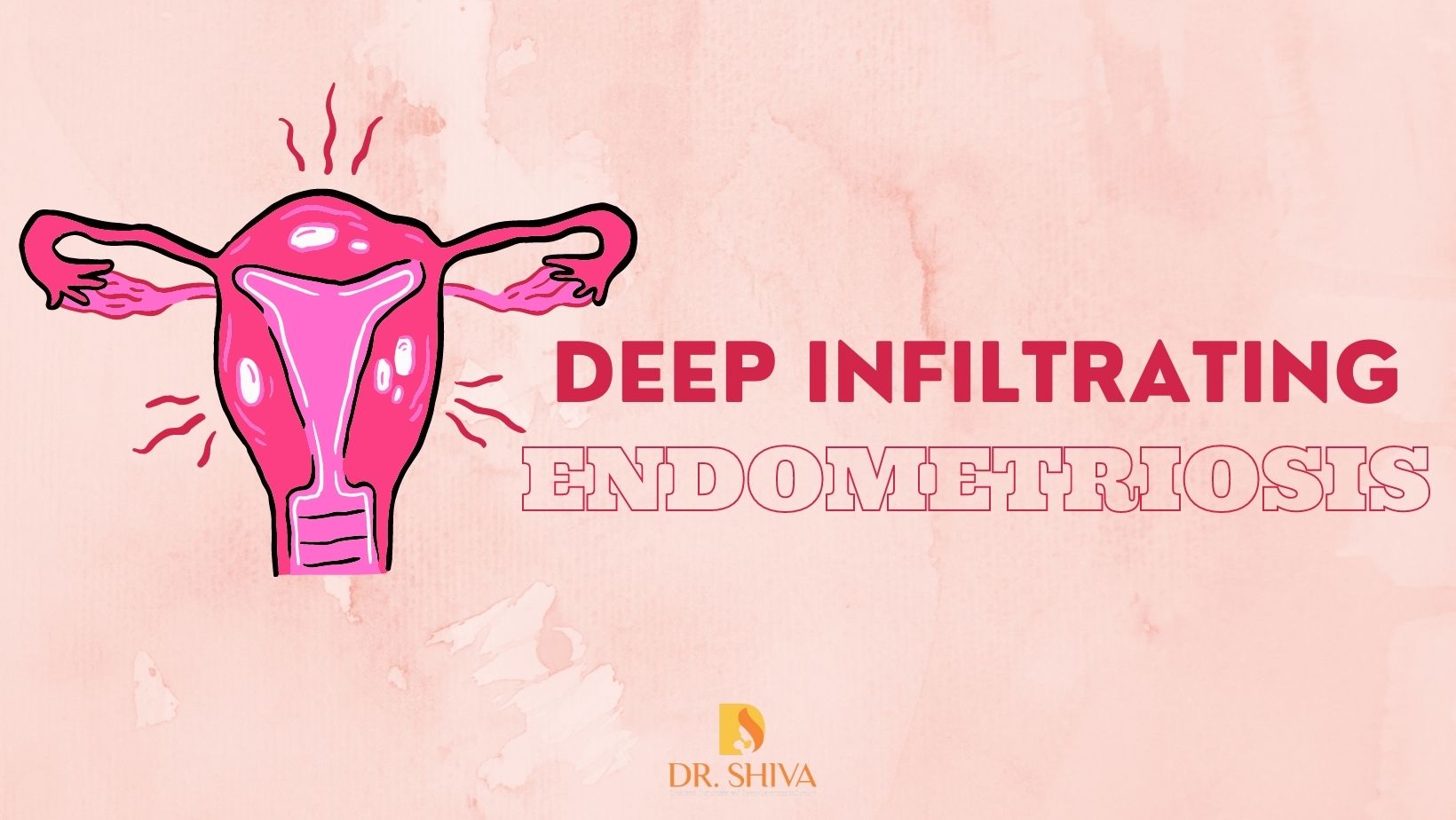
When the endometriosis tissue growing outside the uterus spreads deep inside the tissue of organs close to or inside the pelvic cavity, including the bowel, urinary bladder, intestine, vagina, etc it is called DIE – Deep infiltrating endometriosis. It penetrates more than 5mm into the peritoneal tissues. DIE is rare and usually found only in 1% of women with endometriosis, but the issue is that it may not always respond positively to treatment.

The clumps of scar tissue formed as a healing process after bleeding due to inflammation of endometriosis tissue is called adhesions. These adhesions may bind the organs together.

In this manner, DIE invasion can cause a frozen pelvis, where endometriosis that has progressed deep between organs, locks them together and prevents or restricts their movement. This can cause extreme pain, especially during ovulation, intercourse, and menstrual period.

Symptoms of Deep infiltrating endometriosis

The presence or absence of these symptoms does not indicate the severity of the situation as women with mild symptoms can have a severe form of endometriosis and vice versa too. DIE usually affects areas rich in nerve endings which can cause irritation and pain. The symptoms also vary based on where it has been affected.

-Symptoms of endometriosis in the reproductive tract include pain with mensuration or Dysmenorrhea, pain during intercourse or dyspareunia, and infertility.

-Symptoms for endometriosis in the urinary tract – When the ureter below the pelvic area is affected, it is known as Urinary Tract endometriosis. Some of the symptoms include increased urine frequency, urgency to pass urine, pelvic or lower back pain, burning sensation when passing urine, and presence of blood in the urine. In some cases, the symptoms will not be there, but the ureter will become closed causing a loss of function of the kidney.

-Symptoms for endometriosis in the bowel – Endometriosis in the bowel can cause fusion of the rectum and vagina. This can cause severe pain during intercourse or bowel movements. Abdominal pain, bloating, constipation, diarrhoea, and blood in stool are a few other symptoms. Some do not have any symptoms at all.

Diagnosis for DIE

The doctor will utilize a combination of techniques including pelvic exams, ultrasound scans, MRI scans, laparoscopy, or biopsies to locate and assess the severity of the issue. These diagnostic tools play a crucial role in identifying and understanding the extent of the condition.

Management of Deep infiltrating endometriosis

The treatment approach for deep infiltrating endometriosis (DIE) depends on the issue to be solved or cured, such as pain relief, reduction of endometriosis tissue growth, fertility enhancement, or prevention of recurrence. Hormone treatment may be prescribed to limit estrogen production, which promotes the growth of endometriosis tissue. Surgical interventions, such as removing endometrial tissues, may also be considered. But complete management using medicines or hormone tablets is not possible in most cases.

Laparoscopy to remove the lesions and scar tissue is a common procedure. But it also depends on the magnitude of how much endometriosis has spread.

In severe cases, an incision will be necessary. As the surgery can be complicated it is necessary to have a highly specialized surgeon perform the procedure. This is because an incomplete resection will result in the patient coming again for another surgery. The surgery will provide relief for a few years but there are chances the disease may recur. Treatment via surgical methods also depends on the location of endometrial tissue that needs to be removed.

Urinary tract endometriosis treatment 

The affected part of the ureter needs to be removed and the rest is sewn back. In some cases, the scar tissue only can be removed.

In rare cases when endometriosis has affected a large section of the ureter cutting and re-joining the ureter will not work. Instead, the urinary bladder will have to be moved closer to the ureter and the ureter is inserted into the bladder. During the procedure, a urinary stent will be placed along the length of the ureter from the kidney to the bladder. This stent will be carrying the urine until the ureter heals. The stent will be removed after 6 weeks using a cystoscope.

In cases where endometriosis affects the urinary bladder, the surgical procedure involves the removal of the affected area and suturing the remaining portions together. During the healing process, a catheter will be used for one week.

Bowel Endometriosis Treatment 

If the endometriosis has affected the bowel, depending upon the depth and the size of the nodule of endometriosis, we have to do a shaving of the rectal wall, a discoid excision, or a complete resection of the bowel with an end-to-end anastomosis. There is no need for a colostomy. We will involve the colorectal surgeon; it will be multidisciplinary care.

In cases where the symptoms are not present, and infertility is not an issue the doctors may wait to see what needs to be done.

Dr. Shiva Harikrishnan is one of the best endometriosis specialists in Dubai. She is ranked second in the world and the world’s first female surgeon to have been accredited as a Master Surgeon in Multidisciplinary Endometriosis Care – MSMEC, accredited by the Surgical Review Corporation(SRC), USA. Also accredited as Surgeon of Excellence in Endometriosis Care at Medcare Women & Children Hospital. Watch the video here.

Here is a testimonial by a patient who was suffering from Deep Infiltrating Endometriosis for 15-20 yeasrs and was successfully diagnosed and treated by Dr.Shiva Harikrishnan.

For more details kindly contact us. You may mail to drshivahk@gmail.com or call/Whatsapp  +971557506174.


Cervical and Endometrial polyps

Cervical and Endometrial Polyps

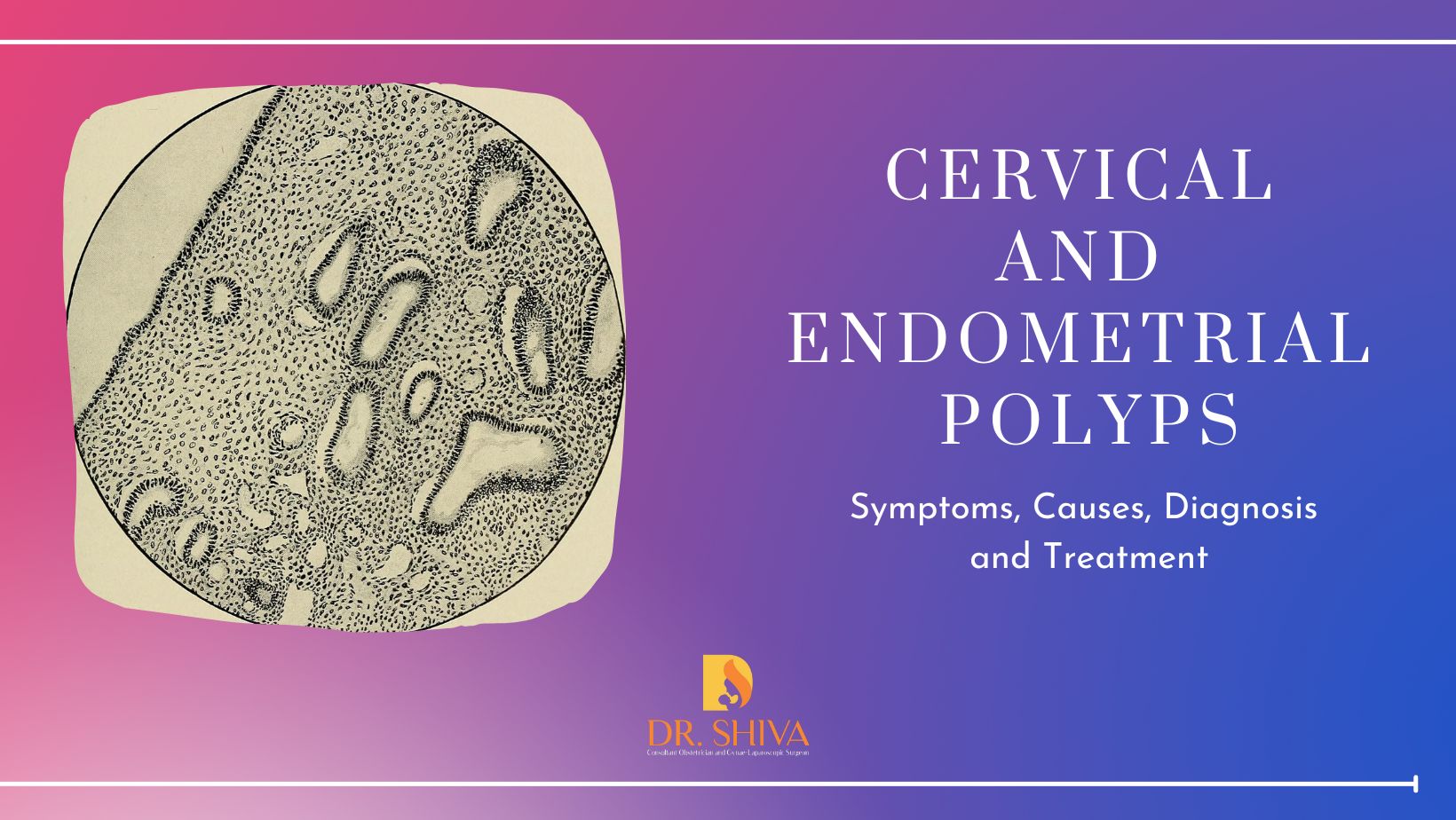
A polyp is a nodule or lump that grows on the inner lining of the organs. An endometrial polyp develops from the endometrial tissue and grows inside the uterine cavity. A cervical polyp originates from the epithelial cells of the cervix and grows on the outside of the uterus, at the top of the vagina. Typically, these polyps are not highly detrimental, primarily causing symptoms. However, there is a slight probability, ranging from 1 to 3%, that they may become cancerous.

Symptoms of having cervical or endometrial polyp

  • Irregular menstrual bleeding.
  • Intermenstrual bleeding.
  • Vaginal bleeding after intercourse
  • Bleeding after menopause.
  • Infertility in some.

Some do not experience any symptoms instead only identified during an ultrasound (endometrial polyp) or pap smear(cervical polyp).

Causes and risk factors

Estrogen levels contribute to the development of endometrial polyps. Having certain medications as a treatment for breast cancer can result in higher estrogen levels. Being obese and high blood pressure are other causes. Infection and chronic inflammation can cause cervical polyps.

Diagnosis 

  • Hysteroscopy – During hysteroscopy, a medical professional inserts a hysteroscope through the cervix into the uterus to view the uterine cavity and identify any polyps. The procedure also allows for the removal of the polyps using the same instrument.
  • Hysterosonography (HSG) – During hysterosonography (HSG), a medical professional injects sterile water into the uterine cavity, causing the uterus to expand. This procedure helps provide a clearer view of the polyps and facilitates their identification.
  • Pelvic Ultrasound – This procedure provides a view of the internal area and helps identify the presence of any polyps.

The polyps can be of two types

  • Symptomatic- in this case, the removal of the polyps will help to resolve the issue caused such as bleeding or discharge.
  • Asymptomatic – Removing asymptomatic cervical polyps and endometrial polyps is necessary to exclude the chances of cancer developing.

Treatment 

While medications may be provided to reduce the symptoms, it is important to note that the effects are temporary. Treatment is the removal of these polyps. Cervical polyps present on the outside of the uterus are comparatively easier to find and remove. Endometrial polyps which are found inside the uterine cavity will require a more detailed procedure. The polyps are removed by scraping or resecting them. Ring forceps are also used to remove the polyps. Some of them may be sent to the laboratory to check if they are benign or cancerous. The treatment is minimally invasive, and, in most cases, local anesthesia is used.

Recovery

The recovery is fast and there are no major cuts or postoperative pain from this surgery other than mild cramping and slight staining. Polyps once removed do not grow back but there are chances for new ones to come.

Can we prevent cervical polyps and endometrial polyps?

Although regular gynecological check-ups and pap smear tests can help detect cervical polyps and endometrial polyps, it is important to note that they cannot be prevented.

Endometrial Hyperplasia

Endometrial Hyperplasia - Thickening of the uterus lining

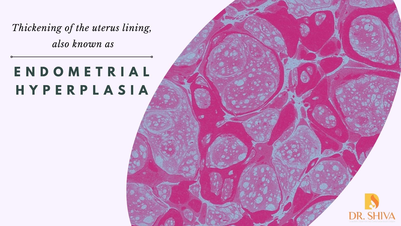
When the uterus lining or endometrium becomes very thick it is known as endometrial hyperplasia. Though it is not cancer, there is a risk of developing endometrial cancer. Endometrial hyperplasia is extremely rare. It can cause unusual or abnormal uterine bleeding.

Causes

The endometrium keeps changing based on the hormone levels. Reduced production of the hormone progesterone and having too much estrogen cause endometrial hyperplasia. The hormone progesterone prepares the uterus for pregnancy and estrogen thickens the endometrium. When conceptions don’t occur the progesterone level drops triggering the uterus to shed the lining. Once it has been shed the next menstrual cycle starts.

Women having endometrial hyperplasia produce a low amount of progesterone due to which the endometrial lining doesn’t shed, and it keeps growing and thickens.

Obesity is another factor that can cause high levels of estrogen because the adipose tissue converts the fat-producing hormones to estrogen.

What conditions lead to endometrial hyperplasia?

  • menopause, when progesterone is not produced as ovulation has stopped
  • the perimenopausal stage when ovulation does not occur regularly
  • when using medicines that act like estrogen that is used for cancer treatment.
  • having diabetes
  • irregular periods especially caused due to PCOS
  • undergoing hormone therapy and not using progesterone

What are the risk factors that can increase the chances of having endometrial hyperplasia?

  • Never been pregnant
  • Obesity
  • Early menstruation or late menopause
  • Having a family history of uterine cancer

Types 

Simple endometrial hyperplasia – this has normal-looking cells which will not be cancerous and may improve without treatment.

Complex endometrial hyperplasia – the abnormal cells may cause precancerous conditions. Treatment is necessary else chances of having cancer will increase.

Symptoms 

  • Heavy bleeding or bleeding that lasts longer than expected.
  • Irregular periods
  • Bleeding after menopause

What are the methods to diagnose for this condition?

Transvaginal Ultrasound – a small device is placed in the vagina and sound waves are send. This is then converted into images which will be examined.

To help identify if the cells are cancerous, it is necessary to take a sample of tissue from the endometrium. Procedures used for this include hysteroscopy and endometrial biopsy.

Treatment 

Progestin can be provided for treatmentThe dosage, duration and method of providing it vary depending upon the age and type of hyperplasia.

If the patient has no plans to have children in the future and if they are not responding properly to the treatment, a hysterectomy may be suggested.

Can it be prevented?

  • If you are having estrogen after menopause, make sure to take progestin too
  • Have birth control pill for irregular periods
  • Maintain a healthy weight.

Complications

Heavy bleeding can make the woman anaemic.

If untreated the chances of cancer developing are around

  • 8% in simple endometrial hyperplasia
  • 30% in complex endometrial hyperplasia.

For more details kindly contact us.